Welcome to SuperLan Chemical Co.,Ltd.
Home > Products > Protein Tyrosine Kinase > VEGFR > Nintedanib

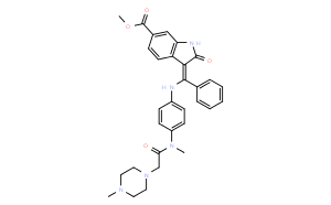
Nintedanib

(CAS No:656247-17-5)
Nintedanib (BIBF 1120) is a potent triple angiokinase inhibitor for VEGFR1/2/3, FGFR1/2/3 and PDGFRα/β with IC50 of 34 nM/13 nM/13 nM, 69 nM/37 nM/108 nM and 59 nM/65 nM in cell-free assays. Phase 3.
CAS No:656247-17-5
Molecular Weight(MW):539.62
Purity:98%+
Specification:500mg;1g;5g;10g;50g;100g
Price and large packaging, please e-mail consultation:info@SuperLan-chem.com
QC Documents
 COA  MSDS  HPLC  NMR
ChemicalInfomation
CAS No: 656247-17-5
Molecular formula(MF) C31H33N5O4
Molecular Weight(MW): 539.62
Alias
Solubility
In vitro DMSO 6 mg/mL (11.11 mM)
Ethanol 3 mg/mL (5.55 mM)
Water <1 mg/mL
In vivo 30% PEG400+0.5% Tween80+5% propylene glycol 30 mg/mL
Biological Activity
Description Nintedanib (BIBF 1120) is a potent triple angiokinase inhibitor for VEGFR1/2/3, FGFR1/2/3 and PDGFRα/β with IC50 of 34 nM/13 nM/13 nM, 69 nM/37 nM/108 nM and 59 nM/65 nM in cell-free assays. Phase 3.
Targets
VEGFR2 [1]
(Cell-free assay)
VEGFR3 [1]
(Cell-free assay)
LCK [1]
(Cell-free assay)
FLT3 [1]
(Cell-free assay)
VEGFR1 [1]
(Cell-free assay)
13 nM 13 nM 16 nM 26 nM 34 nM
In vitro

BIBF1120 inhibits PDGFR kinase activity of PDGFR alpha and PDGFR beta types with IC50 values of 59 nM and 65 nM, respectively. In addition, BIBF1120 suppresses the FGFR subtypes with IC50 of 60 nM, 37 nM and 108 nM for FGFR1, FGFR2, and FGFR3, respectively. BIBF1120 binds to the ATP-binding site in the cleft between the amino and carboxy terminal lobes of the kinase domain. The indolinone scaffold forms two hydrogen bonds with the backbone nitrogen of Cys919 and the backbone carbonyl oxygen of Glu917 in the hinge region. BIBF 1120 inhibits proliferation of PDGF-BB stimulated BRPs with EC50 of 79 nM in cell assays. BIBF1120 at concentrations as low as 100 nM blocks activation of MAPK after stimulation with 5% serum plus PDGF-BB. In cultures of human vascular smooth muscle cells (HUASMC), BIBF1120 prevents PDGF-BB stimulated proliferation with an EC50 of 69 nM. [1]

Cell Data
Cell Lines Assay Type Concentration Incubation Time Formulation Activity Description PMID
SKOV3 Function Assay 5 µM 24 h DMSO induces a significant increase in the promoter activities of E-cad, CDH1, and CDH3 26061747
A549 Function Assay 2/5 μM 24 h DMSO has a general EMT reversal effect  26061747
T24 Function Assay 2/5 μM 24 h DMSO has a general EMT reversal effect  26061747
Mia-Paca2 Function Assay 2/5 μM 24 h DMSO has a general EMT reversal effect  26061747
A549 Function Assay 0.01–5 μM 24 h DMSO induces SFTPD mRNA expression dose dependently 25843005
A549 Function Assay 0.01–5 μM 72 h DMSO enhances SP-D protein expression in a dose-dependent manner at concentrations of up to 5 μM  25843005
A549 Function Assay 5 μM 0-1 h DMSO increases AP-1 activation after 30 min 25843005
Hep3B Cell Viability Assay 0-20 μM 48 h decreases cell viability dose dependently 24657398
HepG2 Cell Viability Assay 0-20 μM 48 h decreases cell viability dose dependently 24657398
PLC5 Cell Viability Assay 0-20 μM 48 h decreases cell viability dose dependently 24657398
HuH7 Cell Viability Assay 0-20 μM 48 h decreases cell viability dose dependently 24657398
SK-Hep1 Cell Viability Assay 0-20 μM 48 h decreases cell viability dose dependently 24657398
Hep3B Apoptosis Assay 0-20 μM 48 h induces cell apoptosis dose dependently 24657398
HepG2 Apoptosis Assay 0-20 μM 48 h induces cell apoptosis dose dependently 24657398
PLC5 Apoptosis Assay 0-20 μM 48 h induces cell apoptosis dose dependently 24657398
HuH7 Apoptosis Assay 0-20 μM 48 h induces cell apoptosis dose dependently 24657398
SK-Hep1 Apoptosis Assay 0-20 μM 48 h induces cell apoptosis dose dependently 24657398
H1703 Growth Inhibition Assay IC50=0.05 μM 23729403

... Click to View More Cell Line Experimental Data

In vivo In all tumor models tested thus far, including human tumor xenografts growing in nude mice and a syngeneic rat tumor model, BIBF1120 is highly active at well-tolerated doses (25-100 mg/kg daily p.o.). This is evident in the magnetic resonance imaging of tumor perfusion after 3 days, reducing vessel density and vessel integrity after 5 days, and profound growth inhibition. [1]