| In vitro |
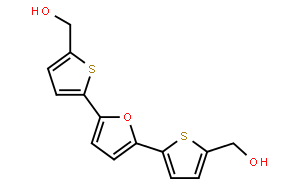
RITA shows a highly selective pattern of differential cytotoxic activity in the tumor cell lines, due to cellular accumulation to the cytosolic (S100) fraction. RITA also inhibits the growth of other renal cell lines including ACHN and UO-31 with IC50 of 13 μM and 37 μM, respectively. [1] RITA (10 nM) causes cell cycle arrest with accumulation of cells at the G2-M phase and induces DNA fragmentation and apoptosis at 100 nM, both with evaluated p53 protein levels. RITA (30 nM) also induces both DNA-protein and DNA-DNA cross-links in A498 cells. Meanwhile RITA has no effects on top1-mediated relaxation of supercoiled SV40 DNA. [2] RITA significantly suppresses the growth of HCT116 cells (97%) but only slightly inhibits the growth of HCT116 TP53-/- cells (13%). RITA is much more efficient at growth suppression in wild-type p53-expressing tumor cell lines than in cell lines lacking p53 and those expressing mutant p53. RITA binds full-length p53 but not glutathione S-transferase (GST) protein or HDM-2 (a key regulator of p53 is strongly supported by the rescue of embryonic lethality of MDM2). RITA blocks p53−HDM-2 interaction and p53 ubiquitination. RITA substantially decreases the amount of HDM-2 that is co-precipitated with p53, although both proteins are upregulated. RITA prevents interactions between the purified GST-p53 and 6XHis-tagged His-HDM-2 proteins. [3] RITA is shown to induce apoptosis by promoting p53Ser46 phosphorylation. [4] RITA induces activation of p53 in conjunction with up-regulation of phosphorylated ASK-1, MKK-4 and c-Jun. RITA induces the activation of JNK signaling. [5] But On the contrary, another results by nuclear magnetic resonance (NMR) show that RITA does not block the formation of the complex between p53 (residues 1-312) and the N-terminal p53-binding domain of MDM2 (residues 1-118), which is highly probable that the binding of RITA requires native conformation of p53. [6]
|

 COA
COA MSDS
MSDS HPLC
HPLC NMR
NMR